
Call/Text for A Consultation Today : CALL/TEXT US: 405-632-5561
Oklahoma's Dental Care Services
Oklahoma's Top Rated Local®
Cavities
Cavities, Decay, Caries
As teeth become exposed to sugar and other acid substances, the enamel of the teeth begins to break down and allow pathogens into the tooth. These pathogens continue to progress to the oxygen supply within the tooth (in the pulp). As you can see from the following x-ray, there is a hint of shadowing that indicates a cavity. The cavity was not evident in the mouth.
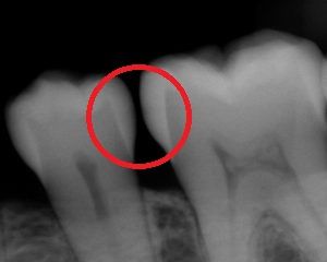
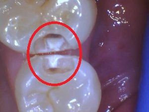
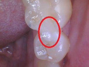
But since the teeth in this x-ray do not touch, this patient was likely trapping food between the teeth causing food impaction which can become painful. In this case, we opted to treat the two teeth. Here’s a pic of the teeth after we drilled out about half of the cavity. Notice how the decay started on the outer surface (widest part of cavity) of the tooth and funnels to the Dentin-Enamel Junction (DEJ). Upon reaching the DEJ, the dentin is softer and more vascular so the pathogens can spread quickly (dark brown spot inside the enamel layer). The following image illustrates the filled cavity with a composite resin filling.
So what’s the harm in letting this cavity remain? It doesn’t hurt so why can’t I just wait?
This decay will continue to progress to the pulp of the tooth where the blood supply is. Once it reaches the pulp, the body starts the inflammation process with no room to swell. The body in turns sees that infected tooth as an infection and must isolate and get rid of it. The body then removes bone around the tooth in an effort to make it loose and fall out. Once the tooth is out, infection is isolated to that small area of the body and not able to grow. The x-ray below shows a tooth that needed to come out. The dark area is infection that is entering the bloodstream. The infection can now spread throughout the body to eventually cause cellulitis.

Please get your teeth cleaned regularly so that we can intercept these cavities before they get worse!
